الملخص الإنجليزي
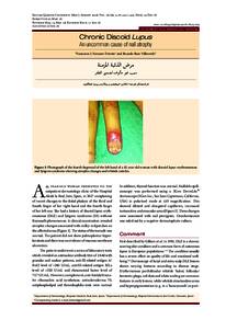
A 45 year-old woman presented to the outpatient dermatology clinic of the Hospital Alcalá la Real, Jaén, Spain, in 2017 complaining of recent changes to the distal phalanx of the third and fourth finger of her right hand and the fourth finger of her left one. She had a history of discoid lupus erythematosus (DLE) and Sjögren syndrome (SS) without Raynaud's phenomenon. A clinical examination revealed atrophic changes associated with milky-red patches on the affected areas. The status of the toenails was normal. The patient did not show palmoplantar hyperkeratosis and there was no evidence of mucous membrane ulceration. The patient underwent a series of laboratory tests which revealed an antinuclear antibody titre of 1/640 with granular and nuclear patterns, anti-SS-related antigen A/Ro52 level of >240 U/mL, anti-SS-related antigen B/La level of >320 U/mL and rheumatoid factor level of 72.7 UI/mL. However, complement, anti-histidyl transfer ribonucleic acid synthetase, antiscleroderma 70, antiphospholipid and thrombophilia tests were normal. In addition, thyroid function was normal. Nailfold capillaroscopy was performed using a 3Gen DermLite® dermascope (3Gen Inc., San Juan Capistrano, California, USA) in polarised mode at x10 magnification. This showed dilated and elongated capillaries, increased tortuosities and avascular areas. These changes were associated with nail pterygium. Onychomycosis was ruled out by a negative dermatophyte culture.