English abstract
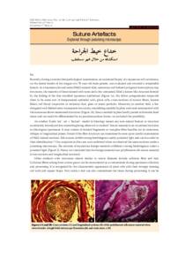
Recently, during a routine histopathological examination, an excisional biopsy of a squamous cell carcinoma,
on the lateral border of the tongue of a 70-year-old male patient, was evaluated and revealed a remarkable
feature. In a haematoxylin and eosin (H&E) stained slide, numerous well defined polygonal isomorphous tiny
structures, the majority of them stained with eosin and a few unstained, filled a lumen-like structure formed
by the folding of the thin stratified squamous epithelium.