وثيقة
Congenital lingual melanotic macule : rare entity in infants.
المعرف
DOI: 10.18295/squmj.2015.15.03.024.
المساهمون
عناوين أخرى
الطاخة الخلقية الصبغية في اللسان : مرض نادر في الأطفال الرضع
الناشر
College of Medicine, Sultan Qaboos University.
ميلادي
2015-08
اللغة
الأنجليزية
الموضوع
الملخص الإنجليزي
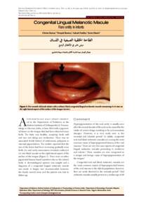
A five-month-old male infant present ed to the Department of Pediatrics at the Madras Institute of Orthopaedics & Trauma tology in Chennai, India, in June 2014 with a pigment ed lesion on the tongue that had been observed since birth. The baby was healthy, accepting feeds well and was not taking any medication. There was no associated family history of melanomas, polyposis or mucosal pigmentation. The mother reported that the size of the lesion had been increasing gradually since birth. An oral cavity examination revealed a solitary 6 x 6 mm black macule on the right lateral aspect of the surface of the tongue. There were no other pigmented lesions found anywhere else on the infant's body. A dermatological opinion was sought and a diagnosis of a congenital lingual melanotic macule was made. A biopsy was recommended; however, the family moved away and the patient was lost to follow-up. Patients with congenital lingual melanotic macules need to be followed up regularly to note any changes in the size, shape or colour of the lesion. Long-term outcomes of patients with congenital lingual melanotic macules are not known, as fewer than ten cases have been reported and the cause of origin of the lesion remains uncertain.6 There is no specific treatment for oral melanotic macules, although many researchers recommend complete excision and histological examination of the lesion.
المجموعة
URL المصدر
zcustom_txt_2
Kumar, Chetan, Sharma, Deepak, Pandita, Aakash, & Shastri, Sweta (2015). Congenital Lingual Melanotic Macule : Rare entity in infants. Sultan Qaboos University Medical Journal, 15 (3), 440–441.
الملخص العربي
قدم رضيع يبلغ من العمر خمسة أشهر إلى قسم طب الأطفال في معهد مدراس لجراحة العظام والرضوض في تشيناي ، الهند ، في يونيو 2014 مع وجود آفة صبغية على اللسان تمت ملاحظتها منذ الولادة. كان الطفل يتمتع بصحة جيدة ، ويتقبل الرضعات جيدًا ولم يكن يتناول أي دواء. لم يكن هناك تاريخ عائلي مرتبط بالأورام الميلانينية أو داء السلائل أو تصبغ الغشاء المخاطي. ذكرت الأم أن حجم الآفة كان يتزايد تدريجياً منذ الولادة. كشف فحص تجويف الفم عن وجود بقعة سوداء منفردة بحجم 6 × 6 مم على الجانب الأيمن من سطح اللسان. لم يتم العثور على آفات مصطبغة أخرى في أي مكان آخر على جسم الرضيع. تم الحصول على رأي طبيب الأمراض الجلدية وتشخيص البقعة الصباغية الخلقية اللسانية. تم التوصية بأخذ خزعة. ومع ذلك ، ابتعدت الأسرة وخسر المريض المتابعة. المرضى الذين يعانون من لطاخات ميلانينية خِلقية يجب متابعتهم بانتظام لملاحظة أي تغيرات في حجم أو شكل أو لون الآفة. النتائج طويلة المدى للمرضى الذين يعانون من اللطاخات الميلانينية الخلقية اللسانية غير معروفة ، حيث تم الإبلاغ عن أقل من عشر حالات ولا يزال سبب الإصابة غير مؤكد. الختان والفحص النسيجي للآفة.
قالب العنصر
مقالات الدوريات
حجم الملف
217.81 كيلوبايت
0
0