Document
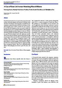
A case of giant left atrium mimicking pleural effusion.
Identifier
DOI 10.5001/omj.2014.82
Contributors
Paul, Rudrajit., Author
Pradhan, Sourav., Author
Choudhury, Partha Sarathi, Author
Das, Shubhabrata., Author
Publisher
Oman Medical Specialty Board.
Gregorian
2014-07
Language
English
English abstract
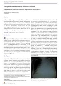
Giant left atrium has become rare in present day owing to decreased incidence with earlier diagnosis and treatment of rheumatic mitral valve disease. Transthoracic ultrasonography is remarkable for clear visualization of giant left atrium with clots. In the absence of clot, ultrasonography picture could be mistaken for aortic aneurysm. In severe left atrial enlargement, chest x-ray may be mistaken for pleural/pulmonary mass or rarely pleural effusion. Ultrasonography and echocardiography may help in diagnosis in such case. A 52 year old female presented with progressive respiratory distress for last two years. She had undergone mitral valve replacement (metallic valve) 24 years back for rheumatic mitral stenosis but for last eight years, the patient did not return for follow-up and had discontinued warfarin. Chest x-ray showed a homogeneous opacity in left hemithorax with obliteration of left costo-phrenic angle suggesting pleural effusion. An ultrasonography of left hemithorax revealed a giant left atrium with spontaneous echogenicity suggesting multiple large non-organized thrombi. There was no pleural/pericardial effusion. Echocardiography also showed a giant left atrium with clots.
Member of
Resource URL
Citation
Santra, Gouranga, Paul, Rudrajit, Pradhan, Sourav, Choudhury, Partha Sarathi, & Das, Shubhabrata (2014). A case of giant left atrium mimicking pleural effusion. Oman Medical Journal, 29 (4), [1-3].
Category
Journal articles
File size
1.14 MB
0
0