Document
Solitary cerebral cysticercus granuloma.
Other titles
ورم حبيبي وحيد لكيسة مذنبة في الدماغ
Publisher
College of Medicine, Sultan Qaboos University.
Gregorian
2011-02
Language
English
English abstract
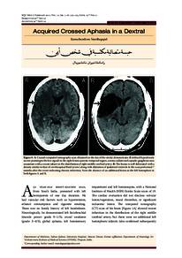
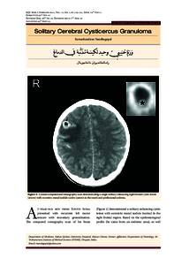
a seven years old boy from South India presented with recurrent left motor seizure with secondary generalisation. The computed tomography scan of his brain demonstrated a solitary enhancing cystic lesion with eccentric mural nodule (scolex) in the right frontal region. Based on the epidemiological profile,as well as the clinical and imaging findings,a diagnosis of solitary cerebral cysticercus granuloma was made. He was treated with a short course of anticysticercal treatment (albendazole), prednisolone
(after excluding spinal and ocular cysticercosis) and phenytoin. Follow-up imaging at 6 months revealed significant improvement.
Member of
Resource URL
Citation
Nandhagopal, Ramachandiran (2011). Solitary cerebral cysticercus granuloma. Sultan Qaboos University Medical Journal, 11(1), 119–121.
Arabic abstract
تعرض صبي يبلغ من العمر سبع سنوات من جنوب الهند لنوبة حركية متكررة في الجانب الأيسر مع تعميم ثانوي. أظهر الفحص بالتصوير المقطعي المحوسب لدماغه وجود آفة كيسية معززة منفردة مع عقدة جدارية غريبة الأطوار (scolex) في المنطقة الأمامية اليمنى. بناءً على الملامح الوبائية ، بالإضافة إلى النتائج السريرية والتصويرية ، تم تشخيص الورم الحبيبي الكيسي الدماغي الانفرادي. تم علاجه بدورة قصيرة من العلاج المضاد للكيسات الكيسية (ألبيندازول) ، بريدنيزولون (بعد استبعاد داء الكيسات المذنبة في العمود الفقري والعين) والفينيتوين. كشف التصوير بالمتابعة في 6 أشهر عن تحسن ملحوظ.
Category
Journal articles
File size
294.66 KB
0
0